US radiology departments are confronting a stark mismatch: imaging demand is projected to climb by 50% by 2030, but the number of trained radiologists is expanding at a far slower rate. Reading images by hand takes time, is vulnerable to fatigue, and produces inconsistent results among different readers. AI-powered medical imaging analysis tools are stepping into this gap, addressing diagnostic imaging, image segmentation, low-dose image enhancement, and regulatory validation. These technologies are changing radiology practice by boosting accuracy, speeding up workflows, and improving patient care.
AI in Radiology and Diagnostic Imaging
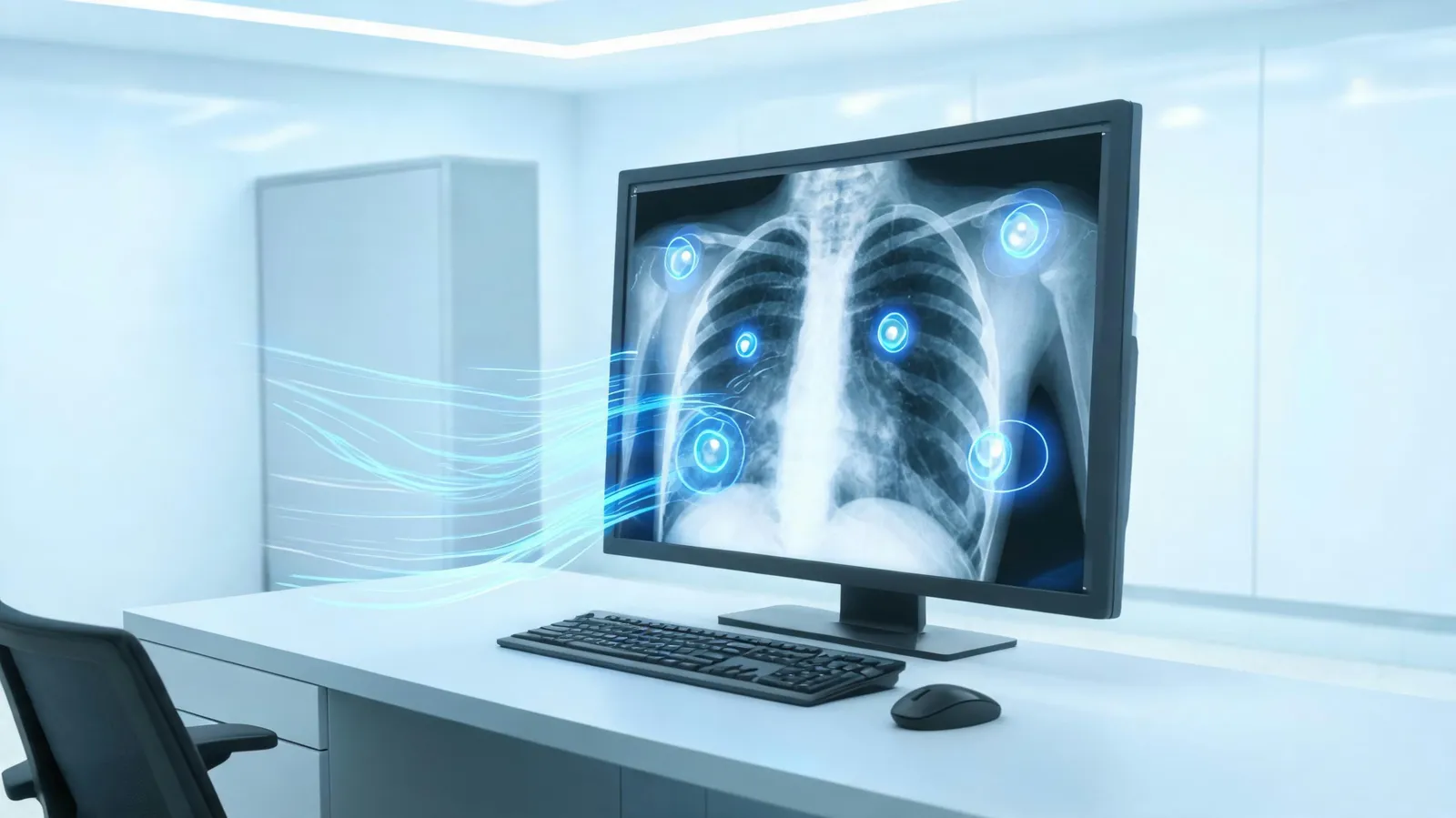
Machine learning algorithms underpin AI-assisted radiology, enabling the detection of abnormalities in X-rays, CT scans, and MRIs. These systems serve as an additional check for radiologists—flagging possible problems, moving urgent cases to the front of the queue, and helping to lower false-negative rates. Multiple studies indicate that AI-assisted mammography screening can cut false negatives by as much as 30%, with particular benefit in dense breast tissue, where human reading is more difficult. In the US, multiple FDA-cleared AI mammography tools are already in clinical use, including systems from Hologic and iCAD. Such tools examine mammograms and mark suspicious areas, helping radiologists prioritize the most critical cases.
Equally critical is how these tools fit into clinical workflows. AI can be integrated directly into Picture Archiving and Communication Systems (PACS) to offer real-time decision support. Research published in 2025 in the Journal of the American College of Radiology reported that AI-assisted triage cut report turnaround time by 22% for critical findings. Radiologist satisfaction also rose: 78% of surveyed radiologists said AI tools helped them handle workload more effectively. Adoption is increasing, but success hinges on smooth integration and clinician trust in the technology.
Image Segmentation for Tumor and Organ Detection
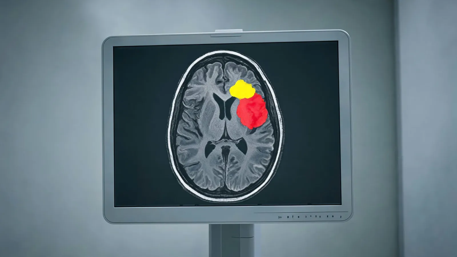
A central component of AI-powered medical imaging analysis is image segmentation. Deep learning models, especially convolutional neural networks (CNNs), can automatically outline tumors and organs with high precision. On public datasets such as BraTS (Brain Tumor Segmentation), leading models reach Dice scores above 0.90 for whole-tumor segmentation—meaning the AI's boundaries overlap expert annotations by more than 90%. In clinical settings, this enables faster and more consistent contouring for radiation therapy planning. For instance, AI-assisted segmentation in brain MRI can reduce contouring time by up to 50% relative to manual methods while also improving consistency between clinicians.
Segmentation tools are also applied outside oncology—for organ detection in abdominal CTs, cardiac MRIs, and prostate MRIs. These uses support surgical guidance, treatment monitoring, and longitudinal studies. The main benefit is reproducibility: AI produces the same output each time, removing the inter-observer variability that can influence diagnosis and treatment planning.
Low-Dose and Portable Imaging Enhancement
Low-dose imaging lowers radiation exposure but often yields noisy images that are harder to read. AI denoising and super-resolution algorithms can improve these images to diagnostic quality. Research shows that AI-enhanced low-dose CT can cut radiation dose by up to 70% without sacrificing diagnostic accuracy. This matters especially for lung cancer screening, where repeat imaging is common. In the US, low-dose CT lung cancer screening is recommended for high-risk individuals, and AI enhancement makes it safer and more accessible.
Portable devices—handheld ultrasound and portable X-ray—are being used more often in point-of-care settings, rural clinics, and emergency response. AI enhancement sharpens image clarity, making these devices more dependable for diagnosis. For example, AI-enhanced portable X-ray has been deployed in rural US clinics to detect pneumonia and fractures in places with limited radiologist access. This technology helps narrow gaps in healthcare access and supports timely treatment decisions.
Regulatory and Clinical Validation of Medical Image AI
Before clinical adoption, AI tools must clear rigorous regulatory review and clinical validation. In the US, the FDA regulates AI-based Software as a Medical Device (SaMD) through pathways such as 510(k) clearance and De Novo classification. The FDA has cleared many AI imaging tools, including those for stroke detection, breast cancer screening, and lung nodule detection. In Europe, the CE mark follows a similar but separate framework. These regulations help ensure AI tools are safe and effective for their intended use.
Clinical validation extends beyond regulatory clearance. Prospective trials and real-world performance monitoring are needed to confirm that AI tools perform across diverse patient populations and clinical settings. Challenges include dataset shift (when the AI encounters data different from its training set), bias, and generalizability. Multi-site validation studies are critical to address these issues. For example, an AI tool trained on data from one hospital may perform poorly in another with different equipment or patient demographics. Ongoing monitoring and updates are necessary to maintain performance.
Conclusion
AI-powered medical imaging analysis tools are reshaping radiology by improving diagnostic imaging, automating segmentation, enabling low-dose imaging, and meeting rigorous validation standards. These advances contribute to better patient outcomes, workflow efficiency, and access to care. As multimodal AI, real-time analysis, and personalized medicine continue to develop, AI's role in radiology will only grow. Healthcare professionals should treat these tools as collaborators rather than replacements, to deliver better care in an increasingly data-driven world.